
Dental Microscope: The Gold Standard That Changes Treatment Quality Forever
In modern dentistry, the line between successful treatment and potential complications is often measured not in millimeters, but in microns. If just a decade ago the microscope was considered the privilege of only elite, narrow-profile specialists, today it is a primary indicator of a clinic's technological maturity and patient care.
"Seeing more" is no longer just a marketing slogan. It is the only objective way to achieve a flawless, predictable, and long-lasting result. Dentistry has moved from the era of tactile, "blind" work to the era of total visual control.
Optical Revolution: Why is the Microscope Indispensable?
A dental microscope is not just a magnifying glass. It is a highly precise, complex optical system that provides magnification of the working field from 2x to 40x (depending on the model) and unparalleled visual control. However, the main secret of the optical revolution lies not only in magnification:
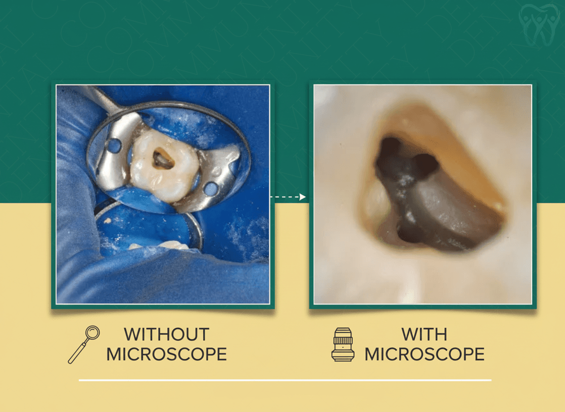
- Coaxial Illumination: Unlike standard dental lights, the microscope's light beam travels in the exact same direction the doctor is looking. This means so-called "dark zones" and shadows disappear. This technology is critically important when working in narrow, deep, and inaccessible canals where standard lighting simply cannot reach.
- Absolute Visual Control: Under the microscope, the doctor can see micro-cracks in the tooth, additional and hidden canals (for example, the 4th canal in upper molars - MB2), and carious lesions at an early stage when they are physically impossible to detect with the naked eye. This protects healthy tissues from excessive preparation (drilling).
Areas of Application: Where Do We See the Maximum Effect?
Although the microscope is used in almost all areas of dentistry, there are fields where it has caused a real breakthrough:
1. Endodontics (Root Canal Treatment)
Without a microscope, endodontics relies heavily on the doctor's intuition and tactile sensation. Using optics makes it possible to:
- Locate and treat canals with the most complex, curved anatomy.
- Safely remove old, low-quality filling materials and safely extract broken instruments.
- Radically increase the success rate of re-endodontics (retreatment) and save "hopeless" teeth.
2. Orthopedics and Aesthetic Restoration
The longevity of an artificial crown, veneer, or filling is directly related to its fit to the tooth structure.
- The microscope ensures ideal preparation with micron precision.
- Fitting the crown margin with maximum accuracy guarantees that bacteria will not get under the structure, and the gums will remain healthy for years.
3. Microsurgery and Periodontics
Using a microscope in surgery means minimal invasion.
- Ultra-fine (micro) sutures are used.
- Soft tissue trauma is minimized, which reduces postoperative pain and ensures the fastest possible rehabilitation for the patient.
The Language of Business: Pros and Return on Investment (ROI)
For clinic management, purchasing a microscope is not just an expense — it is a strategic investment with a clearly measurable return:
- Premium Positioning: The modern patient is informed. When they see high-tech equipment, trust is built. A microscope automatically elevates the clinic to the premium segment and confirms an entirely different standard of quality.
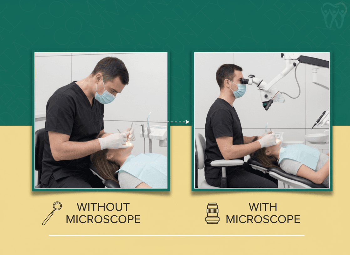
- Doctor's Health (Ergonomics): One of the main problems for dentists is spinal and neck disorders. Working with a microscope forces the doctor to sit in a correct, natural posture, which means preventing occupational diseases and extending the doctor's working capacity for years.
- Minimization of Risks and Costs: Optics sharply reduce the probability of clinical errors. Fewer errors mean fewer complications, avoiding repeated, unplanned patient visits and the obligation to redo work (at the clinic's expense), which directly increases profit.
Real Challenges: What Should We Know?
For the sake of objectivity, it must be said that innovation comes with its own challenges:
- Initial Capital: The microscope and its accompanying instruments require a solid initial financial investment.
- The Learning Curve: Transitioning to new technology takes time. Initially, the doctor's working pace may slow down as they adapt to the new reality of hand-eye coordination. However, once this stage is passed, the achieved precision and the quality of the work performed fully compensate for the time spent.